Knee
What is the Anatomy of the Knee?
The knee is a complex joint made up of different structures - bones, tendons, ligaments, and muscles. They all work together to maintain the knee’s normal function and provide stability to the knee during movement.
Having a well-functioning healthy knee is essential for our mobility and ability to participate in various activities. Understanding the anatomy of the knee enhances your ability to discuss and choose the right treatment procedure for knee problems with your doctor.
Bones of the Knee
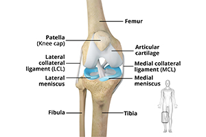
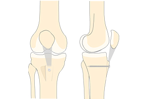
The knee is a hinge joint made up of two bones, the thighbone (femur) and shinbone (tibia). There are two round knobs at the end of the femur called femoral condyles that articulate with the flat surface of the tibia called the tibial plateau. The tibial plateau on the inside of the leg is called the medial tibial plateau and on the outside of the leg, the lateral tibial plateau.
The two femoral condyles form a groove on the front (anterior) side of the knee called the patellofemoral groove. A small bone called the patella sits in this groove and forms the kneecap. It acts as a shield and protects the knee joint from direct trauma.
A fourth bone called the fibula is the other bone of the lower leg. This forms a small joint with the tibia. This joint has very little movement and is not considered a part of the main joint of the knee.
Articular Cartilage and Menisci of the Knee
Movement of the bones causes friction between the articulating surfaces. To reduce this friction, all articulating surfaces involved in the movement are covered with a white, shiny, slippery layer called articular cartilage. The articulating surface of the femoral condyles, tibial plateaus and the back of the patella are covered with this cartilage. The cartilage provides a smooth surface that facilitates easy movement.
To further reduce friction between the articulating surfaces of the bones, the knee joint is lined by a synovial membrane that produces a thick clear fluid called synovial fluid. This fluid lubricates and nourishes the cartilage and bones inside the joint capsule.
Within the knee joint, between the femur and tibia, are two C-shaped cartilaginous structures called menisci. Menisci function to provide stability to the knee by spreading the weight of the upper body across the whole surface of the tibial plateau. The menisci help in load-bearing i.e. it prevents the weight from concentrating onto a small area, which could damage the articular cartilage. The menisci also act as a cushion between the femur and tibia by absorbing the shock produced by activities such as walking, running and jumping.
Ligaments of the Knee
Ligaments are tough bands of tissue that connect one bone to another bone. The ligaments of the knee stabilize the knee joint. There are two important groups of ligaments that hold the bones of the knee joint together, collateral and cruciate ligaments.
Collateral ligaments are present on either side of the knee. They prevent the knee from moving too far during side to side motion. The collateral ligament on the inside is called the medial collateral ligament (MCL) and the collateral ligament on the outside is called the lateral collateral ligament (LCL).
Cruciate ligaments, present inside the knee joint, control the back-and-forth motion of the knee. The cruciate ligament in the front of the knee is called anterior cruciate ligament (ACL) and the cruciate ligament in the back of the knee is called posterior cruciate ligament (PCL).
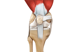
Muscles of the Knee
There are two major muscles in the knee - the quadriceps and the hamstrings, which enable movement of the knee joint. The quadriceps muscles are located in front of the thigh. When the quadriceps muscles contract, the knee straightens. The hamstrings are located at the back of the thigh. When the hamstring muscles contract, the knee bends.
Tendons of the Knee
A tendon is a tissue that attaches a muscle to a bone. The quadriceps muscles of the knee meet just above the patella and attach to it through a tendon called the quadriceps tendon. The patella further attaches to the tibia through a tendon called the patella tendon. The quadriceps muscle, quadriceps tendon, and patellar tendon all work together to straighten the knee. Similarly, the hamstring muscles at the back of the leg are attached to the knee joint with the hamstring tendon.
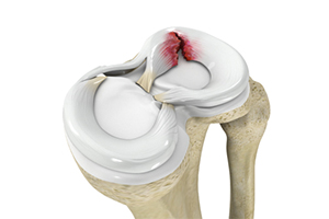
A meniscal tear is a common knee injury in athletes, especially those involved in contact sports. A sudden bend or twist in your knee causes the meniscus to tear. Elderly people are more prone to degenerative meniscal tears as the cartilage wears out and weakens with age.
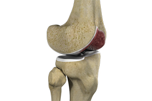
The joint surface is covered by a smooth articular surface that allows pain-free movement in the joint. Arthritis is a general term covering numerous conditions where the joint surface or cartilage wears out. This surface can wear out for several reasons; often the definite cause is not known. Arthritis often affects the knee joint.
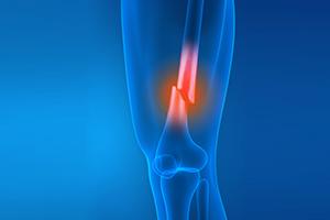
A fracture is a condition in which there is a break in the continuity of the bone. In younger individuals, these fractures are caused by high energy injuries, as from a motor vehicle accident. In older people, the most common cause is a weak and fragile bone.
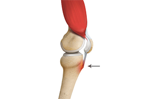
Osgood-Schlatter disease refers to a condition in older children and teenagers caused by excessive stress to the patellar tendon (located below the kneecap). Participants in sports such as soccer, gymnastics, basketball, and distance running are at higher risk for this disease. It is a common knee problem seen in growing adolescents.
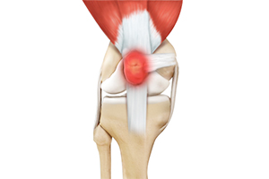
Bursitis refers to the inflammation and swelling of the bursa. Inflammation of the bursa in front of the kneecap (patella) is known as kneecap bursitis or prepatellar bursitis.
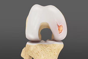
Articular or hyaline cartilage is the tissue lining the surface of the two bones in the knee joint. Cartilage helps the bones move smoothly against each other and can withstand the weight of the body during activities such as running and jumping. Articular cartilage does not have a direct blood supply to it so has little capacity to repair itself.
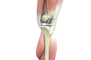
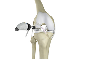
Anterior cruciate ligament (ACL) reconstruction is a surgical procedure to replace a torn or damaged ACL ligament in your knee with a new ACL tissue graft obtained most commonly from your own body (autograft) or in rare cases from a deceased donor (allograft).
Total knee replacement, also called total knee arthroplasty, is a surgical procedure in which the worn out or damaged surfaces of the knee joint are removed and replaced with an artificial prosthesis.
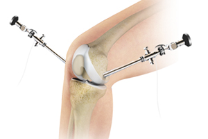
Knee arthroscopy is a common surgical procedure performed using an arthroscope, a viewing instrument, to diagnose or treat a knee problem. It is a relatively safe procedure and you will usually be discharged from the hospital on the same day of surgery.
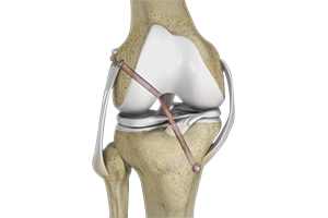
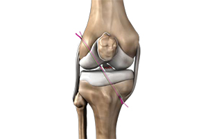
Knee ligament reconstruction is a surgical procedure to repair or replace damaged ligaments of the knee joint. The surgery can be performed using minimally invasive techniques.
Knee surgery is a surgical procedure for the treatment of a knee injury or condition. The procedure involves repairing diseased or damaged structures of the knee joint in order to eliminate pain and restore normal function.
Patella stabilization, also known as patella realignment, is a surgical technique used to stabilize recurrent patellar (knee cap) instability. Patellar instability is a condition characterized by damage to the supporting ligaments that may cause the patella to slip out of its groove either partially (subluxation), or completely (dislocation).
Quadriceps tendon is a thick tissue located at the top of the kneecap. The quadriceps tendon works together with the quadriceps muscles to allow us to straighten our leg. The quadriceps muscles are the muscles located in front of the thigh.
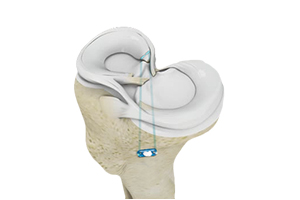
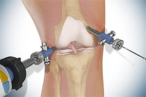
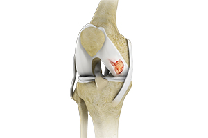
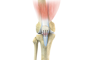
Meniscus root repair is a surgery performed to repair a torn meniscus root. Meniscal repair may be performed either by open surgery under direct vision or minimally invasively using an arthroscope that can be inserted into the knee through a very small key-hole incision to locate and repair the damaged meniscus.
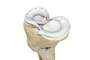
Meniscus repair is an outpatient surgical procedure to repair torn knee cartilage. A variety of minimally invasive procedures are used to repair a torn meniscus, and postoperative protection is required to allow for recovery.
Knee cartilage restoration is a surgical technique to repair damaged articular cartilage in the knee joint by stimulating new growth of cartilage or by transplanting cartilage into areas with defects in order to relieve pain and restore normal function to the knee.
Patellar tendon repair is the surgery performed to reattach the torn tendon to the kneecap and to restore normal function in the affected leg. Patella tendon rupture is the rupture of the tendon that connects the patella (kneecap) to the top portion of the tibia (shinbone). The patellar tendon works together with the quadriceps muscle and the quadriceps tendon to allow your knee to straighten out.
Meniscal surgery is a surgical procedure employed for the treatment of torn or damaged meniscal tissues in the knee. It is mostly performed as a minimally invasive keyhole procedure.
Knee trauma reconstruction is a surgical procedure to repair a soft-tissue injury of the knee such as a torn ligament using a tissue graft or replacing the damaged bony surfaces of the knee joint with an artificial knee joint called a prosthesis. Artificial knee joints are usually made of metal, ceramic or plastic, and consist of the femoral and the tibial components.
