Foot & Ankle
What is the Normal Anatomy of the Foot and Ankle?
The foot and ankle form complex joints that are involved in movement and providing stability and balance to the body. The foot and ankle consist of 26 bones, 33 joints, and many muscles, tendons, and ligaments.
Bones of the Ankle
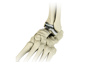
The ankle joint connects the leg with the foot and is composed of three bones: the tibia, fibula, and talus. The tibia or shinbone and fibula or calf bone are bones of the lower leg, which articulate with the talus or ankle bone, enabling up and down movement of the foot.
Three bony bumps present on the ends of the tibia and fibula form parts of the ankle joint:
- The medial malleolus, formed by the tibia, is found on the inside of the ankle.
- The posterior malleolus, also formed by the tibia, is found at the back of the ankle.
- The lateral malleolus, formed by the fibula, is found on the outer aspect of the ankle.
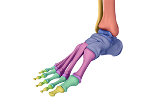
Bones of the Feet
The foot acts as a single functional unit, but can be divided into three parts: the hindfoot, midfoot and forefoot.
The hindfoot forms the ankle and heel, and is made up of the talus bone and calcaneus or heel bone. The heel bone is the largest bone in the foot.
The midfoot connects the hindfoot to the forefoot, and consists of one navicular bone, one cuboid bone, and three cuneiform bones. The navicular bone is found in front of the heel bone, and the cuneiform and cuboid bones are arranged in front of the navicular bone.
These bones are connected to five metatarsal bones of the forefoot that form the arch of the foot for shock absorption while walking or running. The forefoot is also made up of the toes or digits, formed by bones called phalanges - three in each toe, except the big toe, which has only two phalanges. The big toe has two additional tiny round sesamoid bones in the ball of the foot, which helps in upward and downward movements of the toe.
Ankle and Foot Joints
There are 33 joints in the ankle and foot. They include:
- Hinge joints in the ankle, which allow flexion (bending) and extension
- Gliding joints found in the hindfoot, which allow gliding movements
- Condyloid joints found in the forefoot and toes, which allow the flexion (bending) and extension, adduction, and abduction (sideward movement).
The joints of the foot and ankle provide stability and support the weight of your body, helping you to walk or run, and adapt to uneven grounds.
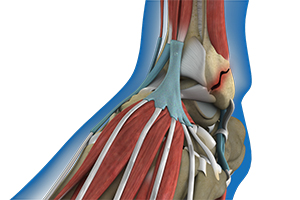
Soft Tissues of the Ankle and Foot
Our feet and ankle bones are held in place and supported by various soft tissues such as cartilage, ligaments, muscles, tendons, and bursae.
The joint surface of all the bones of the ankle and foot are lined by a thin, tough, flexible, and slippery surface called the articular cartilage, which acts as a shock absorber and cushion to reduce friction between the bones. The cartilage is lubricated by synovial fluid, which further enables smooth movement of the bones.
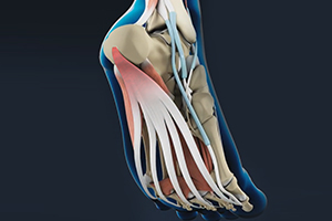
Ligaments are tough rope-like tissue that connect bones to other bones, and hold them in place, providing stability to the joints. The plantar fascia is the largest ligament in the foot, originating from the heel bone to the forefoot, it extends along the lower side of the foot and is involved in maintaining the arch of the foot. The plantar fascia ligament stretches and contracts to provide balance and strength to the foot. Lateral ligaments on the outside of the foot and medial ligaments on the inside of the foot provide stability and allow up and down movement of the foot.
The foot is made up of 20 muscles that help in movement. The main muscles include:
- Anterior tibial muscle, which allows up and down movement of the foot
- Posterior tibial muscle, which supports the arch
- Peroneal tibial muscle, which controls movement on the outside of the ankle
- Extensors, which enable the ankle to raise the toes just before stepping forward
- Flexors, which stabilize the toes against the floor
- Smaller muscles that help the toes to lift and curl
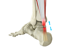
Tendons are soft tissues that connect muscles to bones. The largest and strongest tendon in the foot is the Achilles tendon, present at the back of the lower leg around the heel bone. Other tendons include peroneal and anterior and posterior tibialis.
Bursae are small fluid-filled sacs that decrease friction between tendons and bone or skin. They contain special cells called synovial cells that secrete a lubricating fluid.
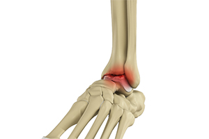
An ankle fracture is a painful condition where there is a break in one or more bones forming the ankle joint. The ankle joint is stabilized by different ligaments and other soft tissues, which may also be injured during an ankle fracture.
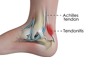
Inflammation of the Achilles tendon is known as Achilles tendonitis or tendinitis. The Achilles tendon is a tough band of fibrous tissue that runs down the back of your lower leg and connects your calf muscle to your heel bone. The tendon is used when you walk, climb, jump, run and stand on your tip toes.
Hallux Rigiditis is a form of degenerative arthritis at the metatarsophalangeal or MTP joint where the base of your big toe attaches to the foot. Arthritis is the inflammation of joints as a result of degeneration of the smooth cartilage that lines the ends of bones in a joint. This degeneration of the cartilages leads to painful rubbing of the bones, swelling, and stiffness in the joints, resulting in restricted movements. Hallux Rigiditis is also called stiff big toe.
Arthritis is the inflammation of joints as a result of degeneration of the smooth cartilage that lines the ends of bones in a joint. This degeneration of the cartilages leads to painful rubbing of the bones, swelling, and stiffness in the joints, resulting in restricted movements. Arthritis in the foot and ankle can occur due to fractures, dislocation, inflammatory disease, or congenital deformity.
Arthritis is the inflammation of joints as a result of degeneration of the smooth cartilage that lines the ends of bones in a joint. This degeneration of the cartilages leads to painful rubbing of the bones, swelling, and stiffness in the joints, resulting in restricted movements. Arthritis in the foot and ankle can occur due to fractures, dislocation, inflammatory disease, or congenital deformity.
A sprain is the stretching or tearing of ligaments. Ligaments connect adjacent bones and provide stability to a joint. An ankle sprain is a common injury that occurs when you suddenly fall or twist the ankle joint, or when you land your foot in an awkward position after a jump. Most commonly, it occurs when you participate in sports, or jump or run on a surface that is irregular.
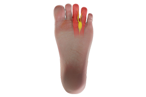
Morton’s neuroma refers to a nerve injury that occurs between the toes, usually the third and fourth toes. This causes pain and thickening of the nerve tissue.
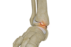
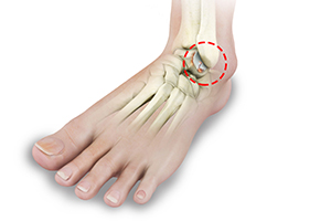
The tibia and the fibula bones of the lower leg join with the talus bone to form the ankle joint. The talus bone is an important bone located between the tibia and fibula and the heel bone (calcaneus). OCL or OCD is the damage to the cartilage and the talus bone of the ankle joint. Usually, the inner or the medial portion of the ankle is affected.
The peroneal tendons run behind the lateral malleolus (the bony protrusion on the outside of the ankle). They connect the peroneal muscles - the peroneus brevis and peroneus longus - to the foot. Peroneal tendon dislocation occurs when either of the tendons slips forward over the lateral malleolus due to a tear in the tendon near its attachment to the bone. When this occurs frequently it can result in inflammation of the tendon causing pain and limiting movement.
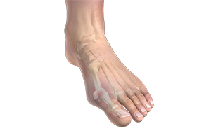
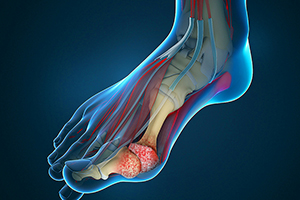
A bunion, also known as hallux valgus, is a bony protuberance that appears on the outer surface of the big toe when it angles toward the adjacent toe. It is an extra bone and a fluid-filled sac that grows at the base of the big toe.
Plantar fasciitis refers to the inflammation of the plantar fascia, a thick band of tissue that is present at the bottom of the foot. It runs from the heel bone to the toes and forms the arch of your foot. Plantar fasciitis is one of the most common causes of heel pain. It is most often seen in middle-aged men and women, but may also occur in those who are constantly on their feet.
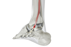
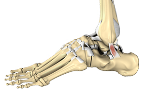
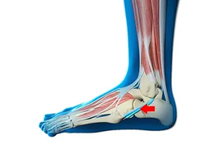
The posterior tibial tendon passes through the ankle to attach the calf muscle with the bones of the midfoot. It provides stability to the arch and supports the foot while walking. Inflammation or a tear of this tendon as a result of injury may cause dysfunction, leading to pain and the development of a flatfoot.
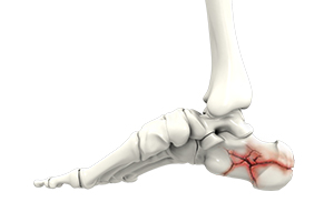
Trauma and repeated stress can cause fractures in the foot. Extreme force is required to fracture the bones in the hindfoot. The most common type of foot fracture is a stress fracture that occurs when repeated activities produce small cracks in the bones.
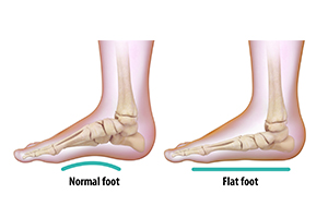
Flatfoot, also known as “fallen arches” or Pes planus, is a deformity in children’s feet where the arch that runs along the sole of the foot collapses to the ground or is not formed at all. Flatfoot is normal in the first few years of life as the arch of the foot usually develops between the age of 3 and 5 years.
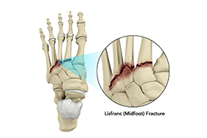
The Lisfranc joint or tarsometatarsal joint refers to the region in the middle of the foot. It is a junction between the tarsal bones (bones in the foot arch) and metatarsal bones (five long bones in the foot). Lisfranc fractures can occur due to a fall from a height or a traumatic motor vehicle accident.
The surgery is usually performed as an outpatient procedure, under a nerve block and sedation. An incision is made at the back of your leg. Your surgeon will stitch the torn tendon back together with strong sutures. Your surgeon may reinforce the Achilles tendon with other tendons depending on the extent of the tear. If the tendon has avulsed or pulled off the heel bone, your surgeon will reattach the tendon to the heel bone.
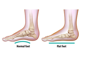
Foot reconstruction is a surgery performed to correct the structures of the foot and restore the natural functionality of the foot that has been lost due to injury or illness. Flatfoot or pes planus is a condition in which the foot does not have a normal arch when standing.
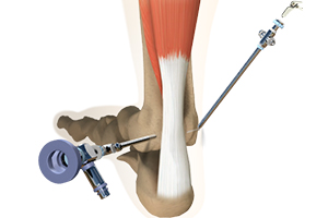
Ankle arthroscopy is a minimally invasive surgical procedure in which an arthroscope, a small, soft, flexible tube with a light and video camera at the end, is inserted into the ankle joint to evaluate and treat a variety of conditions. The camera projects an image of the inside of the joint onto a large monitor, allowing your surgeon to look for any damage, assess the type of injury and repair the problem.
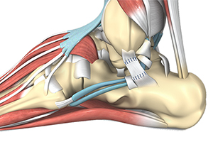
Ankle ligament reconstruction is a surgical procedure typically performed to treat serious sprains or instability in the ankle. Ankle ligament reconstruction aims at restoring stability and function to an ankle that has been compromised by chronic ankle instability or severe ligament injuries. It is effective in repairing torn ligaments, tightening loosened ligaments, and restoring normal stability to the ankle.

Ankle instability is a chronic condition characterized by the recurrent slipping of the outer side of the ankle. Instability is generally noticed during movement of the ankle joint, but can also occur while standing. Ankle instability surgery is performed to treat an unstable ankle and involves the repair or replacement of a torn or stretched ligament.
Ankle joint replacement, also known as total ankle arthroplasty, is a surgical procedure performed to relieve pain and immobility due to severe end-stage arthritis that has not responded to non-surgical treatments. The goal of ankle joint replacement surgery is to eliminate your pain and increase the mobility of your ankle joint.
